Skip to content

Fibroblast-to-Myofibroblast Transition Assay

A unique service that speeds up anti-fibrotic therapeutics development.

Newcells’ FMT assay uses a validated high-throughput system and specialised culture conditions for human primary lung fibroblasts to provide robust data for research on respiratory disorders such as IPF.

Overview

Our FMT-assay improves the understanding of compound efficacy to advance anti-fibrotic drug-discovery programs.

This high throughput assay allows the study of fibroblast activation and collagen deposition in lung fibroblasts following stimulation with  TGF-β.

The specialised set-up enables testing of small molecules and biologics for their ability to prevent fibroblast activation and ECM deposition across a range of concentrations.
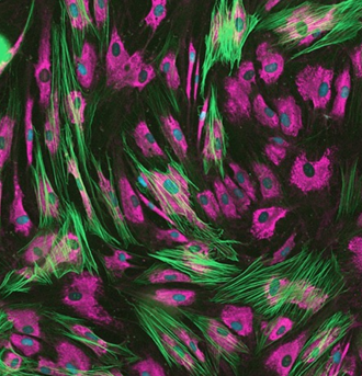
 

Following stimulation with TGF-β1, fibroblasts transition to pre-activated and activated states. Fully activated fibroblasts, shown here, display higher levels of collagen I expression (pink) with an abundance of αSMA incorporated into cellular stress fibres (green). In contrast, under quiescent conditions fibroblasts show a minimal baseline expression of collagen I, which is increased in the ‘pre-activated’ state of proto-myofibroblasts, with an associated diffuse expression of αSMA within the cell cytoplasm.

Newcells’ FMT assay replicates in vitro the changes occurring  following lung injury, mimicking fibroblast activation and matrix deposition. The activation and transition of fibroblasts to myofibroblasts is characterized by the incorporation of α-smooth muscle actin (αSMA) into cellular stress fibres, promoting an increased synthesis and deposition of extracellular matrix (ECM) proteins, such as collagen I, to enable wound repair. Those are accurately quantified in our assay, making it the perfect tool to test novel anti-fibrotic compounds.

High-throughput Imaging
  • Cell number
  • Proliferation rates by (cellular EdU incorporation)
  • Quantification of collagen I expression and deposition
  • Quantification of α-SMA expression

Speak to a Lung Expert


Download our Lung In Vitro Models eBook 

Download E-Book

Newcells' FMT - Assay

Accelerate your lead compound selection by understanding compound efficacy against fibrosis relevant pathology.

1.

Evaluate therapeutic candidates targeting idiopathic pulmonary fibrosis (IPF) and fibrosing lung disease.

2.

Determine compound efficacy using high-throughput imaging and reliable quantification.

3.

Correlate changes in RNA and protein expression

Related reading

Model formats

  • 384-well plate with high-content imaging

Cell Types

  • Primary human lung fibroblasts (HLF)

Species

  • Human (Healthy donor)
  • Human (IPF donor)

Get our latest updates straight to your inbox

About Newcells Biotech

Newcells Biotech provides clients with validated in vitro tools to understand how drugs interact with tissues. We enable the generation of robust and informative data to support critical decisions during any stage of drug-development.

We believe that improving the in vitro models available to drug discovery and development scientists is a key method for increasing the efficiency of in-vitro to in-vivo translation and ultimately to delivering new therapies to patients.

By applying our expertise in iPSCs, primary tissues, a deep understanding of cellular physiology and organoid technology, we have built validated models and assays that have proven to be predictive of how drugs interact with tissues and organs.

Office reception

Innovative company located in Newcastle upon Tyne, in the Northeast of England

Images

A microscope image of a nephron model
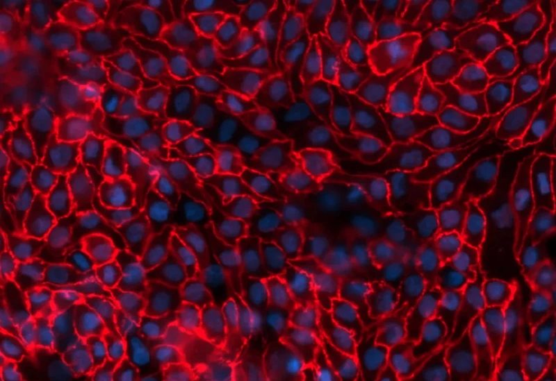
ZO-1 Immunocytochemistry staining of PTCs (red) and Hoescht nuclear staining.
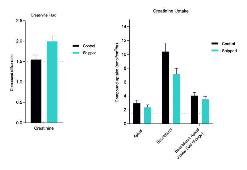
A graph showing different levels of creatine uptake
Functional validation of aProximate™ assay-ready plates for transporter assays Functional validation studies for transcellular flux and uptake of creatinine revealed preferential basolateral-to-apical (JBA) creatinine transport, indicative of normal physiological function. A JBA uptake ratio >1.5 indicates that the cells are functional.

Contact us

Speak to one of our experts about your requirements.

Make an enquiry