Skip to content
Announcement:

Download our latest eBook on lung airway epithelial cell and fibroblast models

96 well plate

Retinal Pigment Epithelium (RPE)

Model formats

  • RPE cells cultured in 24-well Transwell® plates

Cell Types

  • Retinal pigment epithelial (RPE) cells

Species

  • Human (healthy donor)
  • Human (gene-edited)

A functional monolayer in vitro model of retinal pigment epithelial cells generated from human iPSCs for accurate prediction of clinical outcomes

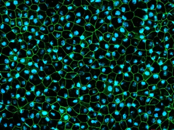
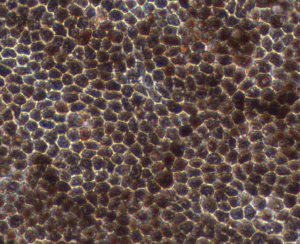
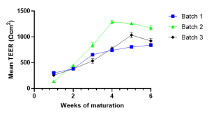
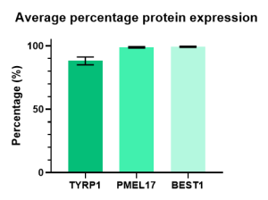
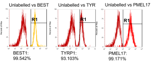
The retinal pigment epithelial (RPE) cell model is composed of a monolayer of RPE cells cultured in 24-well Transwell® plates. RPE characterisation includes: morphology assessment, pigmentation, RPE-specific expression at the protein level (BEST1, TYRP1), the analysis of phagocytosis of photoreceptor outer segments, trans-epithelial resistance (TEER), polarity of apical Pigment Epithelium-Derived Factors (PEDF) and basal vascular endothelial growth factor (VEGF) secretion.

At Newcells, we have extensive in-house experience in retinal in vitro models and can work with you to custom project design and develop assays that suit your requirements. Uniquely, our RPE cells are derived from the same healthy donor iPSCs as our retinal organoids which allows combined assessment of both RPE and neurosensory retina using cells from the same genetic background. We are able to generate RPE cells from both our own and customer-supplied iPSCs to suit various project needs. The model format allows for flexibility in dosing and analytical readouts, including functional assessment of the cells.

Available analytical readouts

  • Immunofluorescence
  • mRNA quantification by RT-qPCR
  • Transcriptomics by single-cell RNA sequencing
  • Growth factor (VEGF, PEDF) secretion
  • Flow cytometry
  • Electron microscopy
Make an enquiry

Don't miss out on our latest innovations: follow us on Linkedin

A microscope image
RPE cells displaying cobblestone morphology. Cells were immunolabeled with tight-junction ZO-1 marker (shown in green) and co-stained with nuclei marker, Hoechst (shown in blue).

Origin

Healthy donor

Outsourced

We do the work

Characteristics

Functional RPE

Retinal Pigment Epithelium Model Highlights

Accelerate your lead compound selection by understanding their mode of action in functional retinal tissue

1.

Mature and functional RPE model

2.

Well-characterised and reproducible

3.

Rapid evaluation of gene therapy vectors

Applications for retinal pigment epithelial cells Close Open
  1. Gene therapy including in vitro viral vector evaluation
  2. Disease modelling
  3. Investigational drug safety and efficacy
Role of RPE in the retina Close Open

The retinal pigment epithelium (RPE) is a single layer of hexagonal-shaped cells that is located between the photoreceptors of the retina and the blood vessels of the choroid, at the back of the eye. The RPE cell layer is critical to vision. The RPE reduces light damage in the eye, regulates the movement of nutrients and waste products in and out of the retina and maintains blood vessels by secreting growth factors. RPE is also responsible for the turnover of photoreceptors.

The RPE is crucial to vision, thus it is important to assess how it is affected by new therapeutic drugs, a task that can be performed in vitro.

Retinal pigment epithelial cells characterisation Close Open

In vitro retinal pigment epithelial cells are generated from human iPSCs to form a 2D monolayer. These RPE cells display typical cobblestone morphology and are pigmented.

Hexagonal morphology and pigmentation typically observed in mature RPE cells

The in vitro model closely recapitulates key functions found in vivo, including formation of tight barriers and phagocytosis of photoreceptor outer segments.

TEER measurements were carried out starting 7 days after seeding on the inserts. RPE reach maturation 4-6 weeks post-isolation when TEER starts to plateau.

Flow cytometry data illustrates a highly pure, differentiated population as demonstrated by high levels of expression of RPE markers BEST1, TYRP1 and PMEL17

Expression of RPE markers, TYRP1, PMEL17 and BEST1
Protein expression of RPE cells using flow cytometry after TEER plateaued. PMEL17 – a protein which is expressed in melanosome precursors (>95% expression). TYRP1 – expressed in mature RPE and is located in melanosomes (>85% expression). BEST1 – is a Ca2+-regulated chloride channel and is a critical for normal phagocytotic function (>98% expression)
Retinal pigment epithelial cells description Close Open
  1. Cell types: RPE cells
  2. Format: Monolayer, cultured in 24-well Transwell® plates
  3. Main characteristics: Cobblestone morphology, pigmentation, express RPE cells biomarkers e.g., BEST1, TYRP1 and RPE65
  4. Other characteristics: Formation of tight barrier and ability to phagocytose photoreceptor outer segments

Outsource your experiments to us

Interested in our RPE model? Speak to one of our experts about your requirements.

Make an enquiry

Get our latest updates straight to your inbox