Latest Publication: Read our teams’ latest paper in Antibiotics, showing reduced nephrotoxicity of a novel formulation of polymyxin B compared to the clinical formulation using a validated primary human proximal tubule cell model.
A robust, physiologically relevant air-liquid interface (ALI) lung model of the small airways to enable scientific research and drug discovery
Newcells Biotech’s human small airway epithelial cell (SAEC) air-liquid interface model closely recapitulates the epithelial physiology of the lung. Derived from differentiated small airway basal cells, our SAEC model comprises the main epithelial cell types namely basal, club (Clara), goblet and ciliated cells. With an established epithelial barrier, active mucus production and functional cilia, our air-liquid interface (ALI) SAEC model is an invaluable tool for drug discovery for the study of lung physiology, drug toxicity and response to epithelial damage.
We currently offer a range of analytical readouts in line with project requirements, example readouts include;
Don't miss out on our latest innovations: follow us on Linkedin
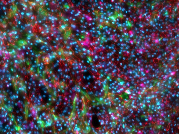
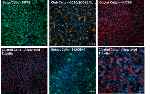
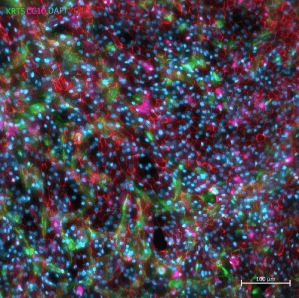
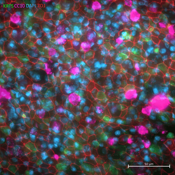
Small Airway Epithelial Cells immunostained to detect for Basal cell (KRT5, green), Club cell (CC10/SCGB1A1, pink) and Junctional protein (ZO1, red) markers.
Human primary
low passage cells
Three human donors
Mimics in vivo tissue
air-liquid interface
Human lung small airway epithelial cell (SAEC) model
A robust, physiologically relevant air-liquid interface (ALI) lung model of the small airways to enable research and drug discovery
1.
Physiologically relevant air-liquid interface (ALI) small airway model
2.
Contains all key lung epithelial cell types
3.
Testing of compounds toxicity on the small airways
Immortalised cell lines and primary cells derived from animals models are commonly used, however, the data they generate lack relevance to human physiology. In order to recapitulate the pseudostratified mucociliary phenotype observed in vivo, human lung small airway epithelial cells need be cultured at the air-liquid interface.
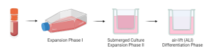
For our ALI-SAEC model, primary human small airway basal cells are expanded in culture flasks before being seeded onto permeable Thincert supports containing culture medium in both the apical and basal compartments. The basal cells are then expanded until confluent prior to being subjected to “air-lift”, i.e. with medium only supplied to the basal chamber to create the air-liquid interface. This configuration mimics the conditions found in human and drives differentiation towards a mucociliary phenotype, forming a 3D pseudostratified epithelium containing club cells in greater abundance than goblet cells. Full differentiation takes 21-28 days and cultures are used from 21-days onwards.
Expression of Junctional Genes & Proteins
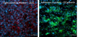
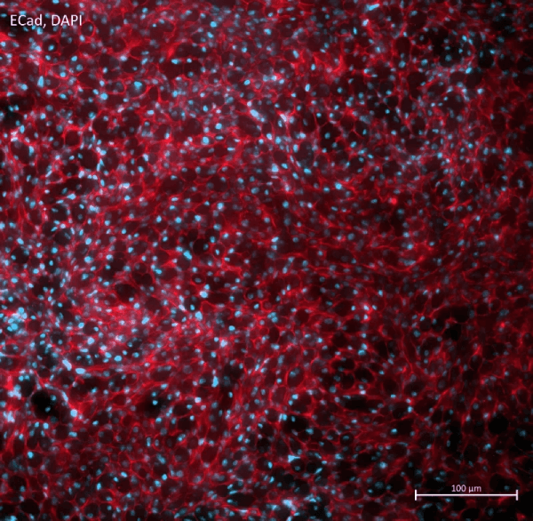
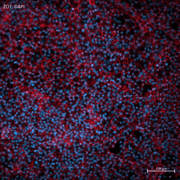
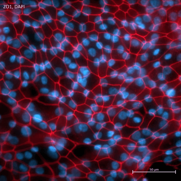
This model is further characterised by the development of a functional epithelial barrier, as indicated by the expression of tight junction proteins and the development of high transepithelial electrical resistance (TEER) as shown by staining of the junctional proteins ZO1 and E-Cadherin. ZO1 and E-Cadherin protein expression, in conjunction with TEER data, confirms cells are polarised and successfully differentiated. Gene expression levels of junctional genes (TJP1, JAM-A, CDH1) can also be tracked throughout the differentiation period.
SAEC cell culture was immunostained for the tight junction marker ZO1 and the adherens junction marker ECadherin
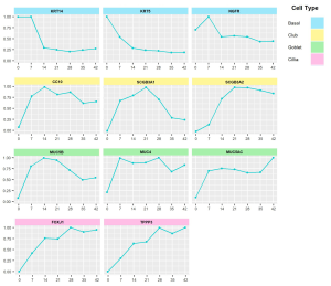
Cell-Specific Gene Expression
As the basal cells differentiate, we quantify the expression of cell-specific genes as an indirect assessment of basal cell differentiation by qPCR. This also confirm the presence of all relevant cell types following 21 days in culture.
Cell-Specific Protein Expression
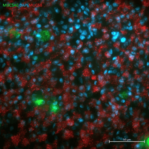
MUC5B and MUC5AC are mucin proteins expressed by goblet cells in both the upper and small airway, however their ratio differs depending on the location. MUC5B is predominantly expressed in the small airways. Validation data collated from the SAEC model suggests a higher expression of MUC5B than MUC5AC, similar to the expression pattern observed in vivo.
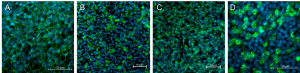
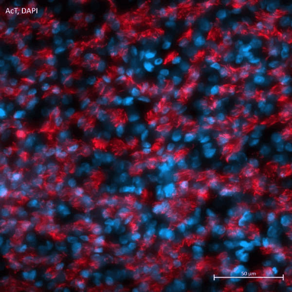
Newcells’ small airway epithelial cell model expresses key cell specific proteins. (A) ZO1 staining shows presence of epithelial tight junctions. Presence of club cells (B), goblet cells (C) and ciliated epithelial cells (D) as indicated by CC10, MUC5B and acetylated tubulin (AcT) expression.
Immunostaining of SAEC model for cell-specific proteins
Brightfield recording of fully differentiated Small Airway Epithelial Cells in air-liquid interface (ALI) cell culture with functional beating ciliary movements.
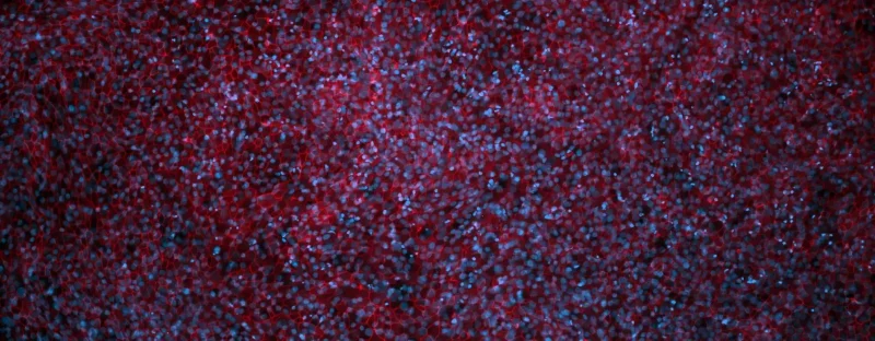
Small Airway Epithelial Cells immunostained to detect Acetylated Tubulin (AcT), a marker for Ciliated cells
Small Airway Epithelial Cells immunostained to detect junctional protein E-Cadherin, a marker for Adherens Junctions
Small Airway Epithelial Cells immunostained to detect junctional protein ZO1, a marker for Tight Junctions
Small Airway Epithelial Cells immunostained to detect junctional protein ZO1, a marker for Tight Junctions
Small Airway Epithelial Cells immunostained to detect junctional protein ZO1, a marker for Tight Junctions
Small Airway Epithelial Cells immunostained to detect mucin proteins MUC5AC (green) and MUC5B (red), which are markers for Goblet cells.
Small Airway Epithelial Cells immunostained to detect for Basal cell (KRT5, green), Club cell (CC10/SCGB1A1, pink) and Junctional protein (ZO1, red) markers.
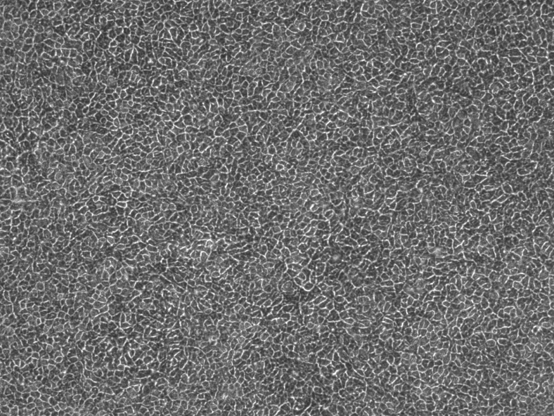
Brightfield Image of Small Airway Epithelial Cells in air-liquid interface (ALI) cell culture.
Small Airway Epithelial Cells immunostained to detect for Basal cell (KRT5, green), Club cell (CC10/SCGB1A1, pink) and Junctional protein (ZO1, red) markers.
How to use our primary small airway epithelial cell model?