Skip to content
An illustration of a retina looking to the left

Advanced Retinal Models –
Revolutionising Retinal Research

Accelerate your retinal research today with our unparalleled models tailored for discovery, innovation, and clinical translation. Contact us now to learn more!

Our retina models have been engineered by experts in iPSCs to mimic human embryonic development and human physiology. The models and assays  have been characterised and rigorously validated to generate reliable comparative data predictive of human responses
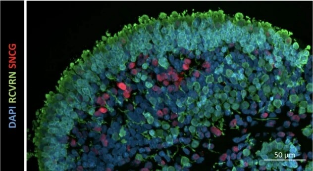
Retinal Organoids

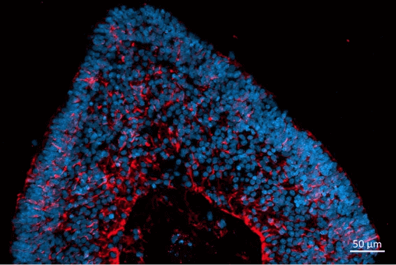
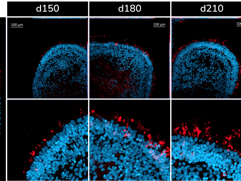
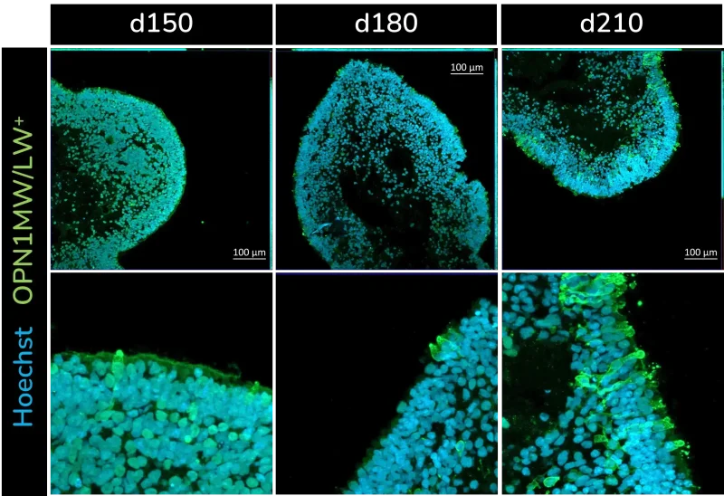
These light-responsive organoids mimic the intricate structure of the human retina, including laminar cell organisation and functional outer photoreceptor segments that respond to light stimuli. They serve as an invaluable tool for advancing science and clinical applications.

Cone photoreceptor cells labelled with anti-Opsin (Red/Green) antibody.

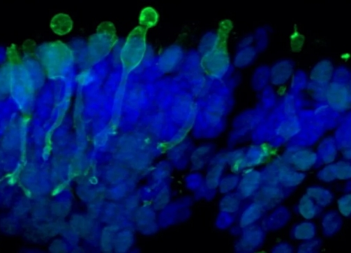
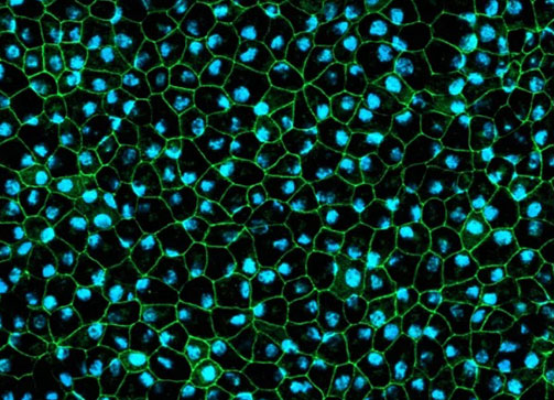
Retinal Pigment Epithelium (RPE)

A functional 2D model of retinal pigment epithelial cells generated from human iPSCs recapitulating phagocytosis of photoreceptor outer segments. The RPE cells are pigmented and displays typical cobblestone morphology.

RPE cells displaying cobblestone morphology. Cells were immunolabeled with tight-junction ZO-1 marker (shown in green) and co-stained with nuclei marker, Hoechst (shown in blue).

Bring your ocular research to the next level with our state-of-the-art retinal organoid (RO) and retinal pigment epithelium (RPE) models, engineered for unmatched precision in predicting clinical outcomes.

Speak to a Retina Model Expert


Download Our Retinal Models E-Book

The front cover of the retinal organoid ebook

Download E-Book

Why use validated retina tissue models?

1.

Accurately mimic physiology of the human retina

2.

Well-controlled production process

3.

Readily available

Related reading

Products

  • Fresh retinal organoids
  • Retinal organoid cell pellets
  • Frozen sections of retinal organoids
  • Standard offering at Day 150 (D150)
  • Customisable time points available D30, D60, D90, D120, D180, D210

Cell Types

  • Rod and cone photoreceptors
  • Retinal ganglion cells
  • Bipolar cells
  • Horizontal cells
  • Amacrine cells
  • Müller glial cells

Origin

  • Human iPSCs (healthy donor)

Get our latest updates straight to your inbox

About Newcells Biotech

Newcells Biotech provides clients with validated in vitro tools to understand how drugs interact with tissues. We enable the generation of robust and informative data to support critical decisions during any stage of drug-development.

We believe that improving the in vitro models available to drug discovery and development scientists is a key method for increasing the efficiency of in-vitro to in-vivo translation and ultimately to delivering new therapies to patients.

By applying our expertise in iPSCs, primary tissues, a deep understanding of cellular physiology and organoid technology, we have built validated models and assays that have proven to be predictive of how drugs interact with tissues and organs.

Office reception

Young innovative company located in Newcastle upon Tyne, in the Northeast of England

Images

RPE cells displaying cobblestone morphology. Cells were immunolabeled with tight-junction ZO-1 marker (shown in green) and co-stained with nuclei marker, Hoechst (shown in blue).
Cone photoreceptor cells labelled with anti-Opsin (Red/Green) antibody.
Localization and distribution of Müller glia cells (CRALBP, red) in retinal organoids. Nuclear DAPI staining (blue).
Presence of rod photoreceptors (RHO+) at different stages of development in iPSC-derived retinal organoids. ‘d’ refers to ‘days of differentiation’
Presence of green and red cone photoreceptors (OPN1MW/LW+) at different stages of development in iPSC-derived retinal organoids. ‘d’ refers to ‘days of differentiation’.
Localization and distribution of photoreceptors (RCVRN, green) and retinal ganglion cells (SNCG, red) in retinal organoids at d150. Nuclear DAPI staining (blue)

Contact us

Speak to one of our experts about your requirements.

Make an enquiry