Skip to content
Announcement:

Join us for our next webinar on Wednesday 7th May 2025, where our 2 panelists, Dr Rodrigues & Dr Katz, will discuss the real impact of in vitro kidney models in producing higher quality data and ultimately safer drugs.

Retina Efficacy Study

Predictive efficacy testing on human retinal models

Accelerate your drug development using human retinal organoids and RPE models

1.

Recapitulate the architecture & function of the human retina

2.

Rapid evaluation of novel compounds’ efficacy

3.

Accelerate drug discovery

Efficacy testing of novel therapies on predictive human in vitro retinal models

Retinal compound efficacy study: this service evaluates how a compound or viral vector affects structure and function of retinal cellular models.

The in vitro drug efficacy study allows rapid evaluation of the efficacy of your drug on high quality healthy and/or diseased human IPSC-derived retinal organoids and/or RPE models. The models are highly representative of the structure and function of human retinal tissues and can also be generated from patients’ iPSCs or gene edited iPSC lines, thus facilitating predictive advanced in vitro testing of new therapeutics and accelerating lead compound selection.

The assays can be carried out at different time points of organoid differentiation and will provide predictive data to allow for confident decision making to progress your new drug or submit an IND.


Service outputs

  • Rapid retinal efficacy evaluation of compounds in retinal organoids and RPE
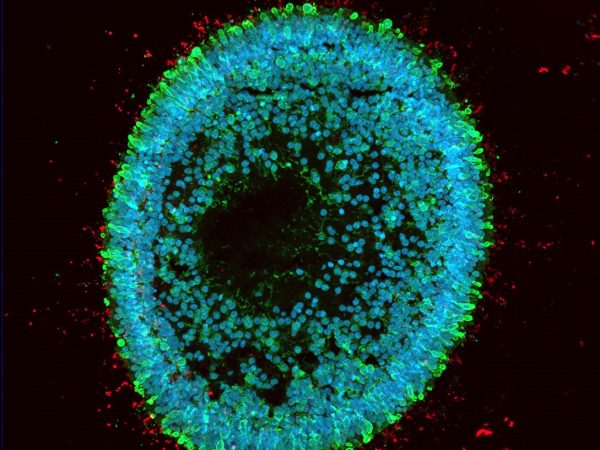
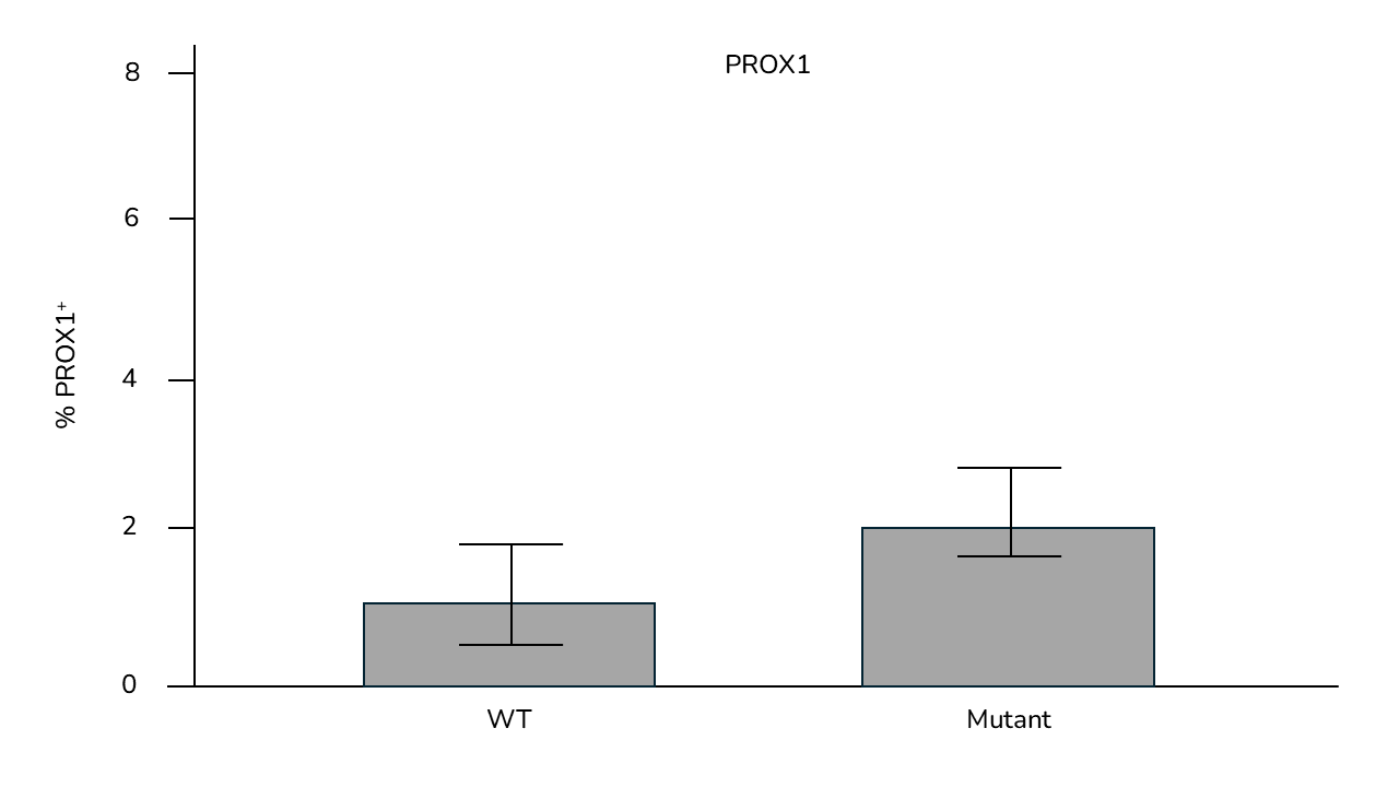
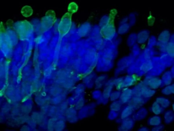
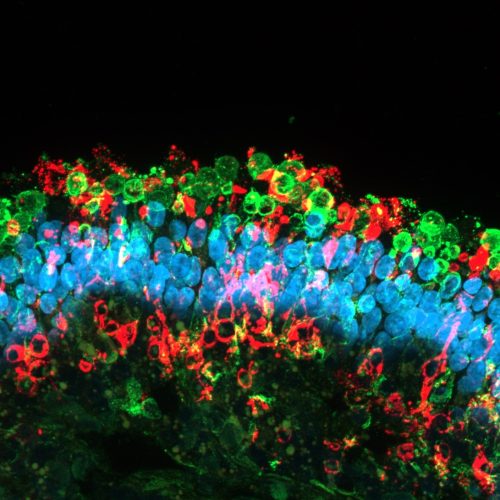
  • Quantitative and qualitative analysis of key markers expression by gene expression analysis and immunofluorescence on frozen retinal organoid sections
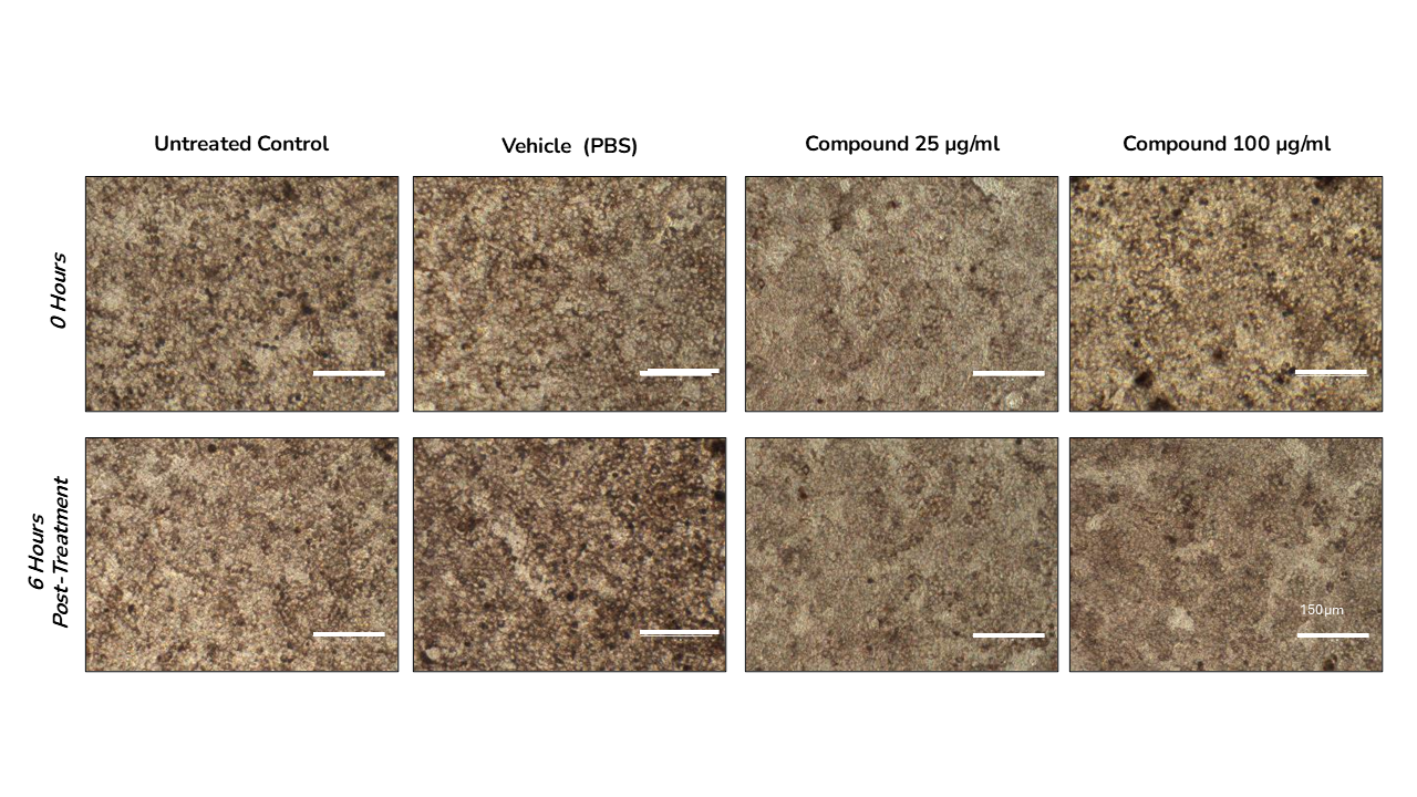
  • Time points comparisons
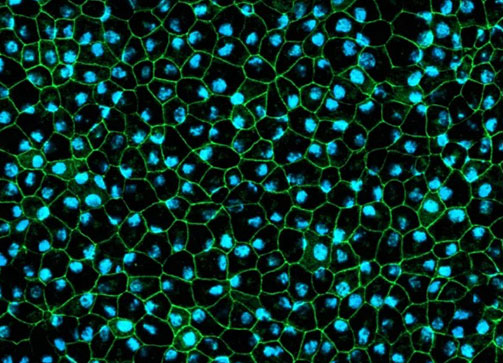
  • Compound effect on PRE barrier, phagocytic integrity, viability and ultrastructure
Make an enquiry Download fact sheet Download eBook

Retinal therapy efficacy study

96-well format

Rapid and predictive

Assays

  • Retina efficacy study: Cell morphology and cell viability; key markers analysis and functional readouts

Models

  • Human retinal organoids (RO)
  • Human retina pigment epithelium (RPE)
  • Models are derived from WT iPSCs or Patient/gene-edited iPSCs supplied by the client

Timeline

  • 2-6 months
Service overview Close Open

Newcells provides retinal efficacy study service using our highly reproducible iPSC-derived human models (retinal organoids and RPE) to provide you with predictive data

Retinal compound efficacy studies are carried out on our highly reproducible iPSC-derived human models (retinal organoids and RPE) to provide clients with predictive data for clinical translation and to be included in regulatory applications.

With a streamlined manufacturing process releasing regular supply of highly reproducible tissue, projects timelines are usually short with studies generally completed within two months. The predictive data generated by our scientific experts will therefore allow you to rapidly assess the efficacy of your drugs to confidently guide key decision-making steps during lead compound identification, drug development or regulatory submissions.

An typical retinal compound efficacy study includes a set of advanced in vitro assays to assess cell morphology and cell viability, photoreceptor functionality and degeneration as well as analysis of key marker expression and localisation to test the efficacy of novel drug compounds and viral vectors such as AAV.

Example 1: Testing the effect of a novel therapeutic on retinal organoids Close Open

The effect of a compound was assessed in a retinal organoid model containing a specific mutation found in an ocular disease to determine how it affected the different cell types present in the retinal organoid model.

Quantification graphs of horizontal cells marker PROX1 following treatment with a novel compound; Statistical analysis performed by unpaired t-test (p< 0.01). Data represents mean ± SEM, n=6.

Disease model organoids had significantly higher expression of horizontal cell marker (PROX1) and rod cell marker (RHO) compared to wild type organoids at day 180 of differentiation following treatment with the novel compound. Other cell-specific expression markers for bipolar cells, amacrine, muller glial and astrocytes cells were not affected by treatment.

Example 2: Assessment of Retinal Pigment Epithelial barrier integrity upon drug treatment Close Open

The retina efficacy service on the RPE model assessed the effect of a compound on the barrier properties of RPE. The compound was tested for its ability to disrupt the epithelial barrier such as layer integrity, permeability and cell viability.

Morphological changes of the epithelium were assessed through brightfield imaging.

Representative brightfield imaging (x20 magnification) of RPE cells before and after 6 hours of treatment with 25 and 100 µg/ml of test compound. Controls include media only (untreated control) and vehicle control (PBS). All the images were acquired with the same magnification, using consistent exposures across all conditions.

No notable morphological changes were observed after 6 h of treatment. TEER measurement showed no significant differences upon treatment with the chosen compound, confirming that the tested compound does not significantly affect the RPE barrier integrity.

Models to choose from for this service

Retinal organoids

The human retinal organoids are iPSC-derived microtissue models. They recapitulate the complex structure of the human retina with laminar cell organisation mimicking embryonic development. They contain the outer photoreceptor segment of the retina that responds to light.

A microscope image of retinal organoids
Cone photoreceptor cells labelled with anti-Opsin (Red/Green) antibody.
Find out more

Retinal Pigment Epithelium

A functional human 2D in vitro model of retinal pigment epithelial cells generated from human iPSCs recapitulating phagocytosis of photoreceptor outer segments. The RPE cells are pigmented and displays typical cobblestone morphology making them a complex human in vitro model able to produce predictive translational data.

RPE cells displaying cobblestone morphology. Cells were immunolabeled with tight-junction ZO-1 marker (shown in green) and co-stained with nuclei marker, Hoechst (shown in blue).
RPE cells displaying cobblestone morphology. Cells were immunolabeled with tight-junction marker ZO-1 (shown in green) and co-stained with nuclei marker Hoechst (shown in blue).
Read more

CONTACT US

Are you interested in this service? Speak to one of our experts about your requirements.

Make an enquiry

Get our latest updates straight to your inbox

Book a Call