Accelerate your lead compound selection with predictive preclinical safety testing and mode of action investigations in functional human-derived retinal tissue
1.
Complex functional human retina model
2.
Well-characterised and reproducible
3.
Ready-to-use
Light responsive human retinal organoids for accurate prediction of clinical outcomes
The Newcells retinal organoids are microtissue Microphysiological Systems (MPS) that recapitulate the complex structure of the human retina with polarised laminar cell organisation mimicking embryonic retina development. These human iPSC derived advanced retinal organoids systems in particular contain the light responsive outer photoreceptor segment of the retina.
Invitro retinal organoid differentiation follows the embryonic development timeline, spanning 150 to 210 days. The temporal order of retinogenesis is comparable to in vivo, recapitulating critical features of foetal retinal tissue including the laminar organisation of cell types.
Retinal organoids are microtissues that include retinal ganglion cells, horizontal cells, amacrine cells and photoreceptors (including cone and rod photoreceptors). Depending on the cell type of interest, the retinal organoids can be used at different stages of development (usually between D60 and D210). For example, retinal ganglion cells are more prevalent at D60 whereas the photoreceptors have a peak expression at later time points.
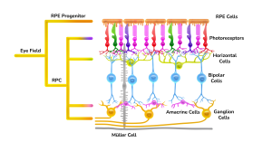
Schematic representation of the retina
The differentiation process is fully characterised and monitored through the analysis of biomarkers specific for each cell type. For example, for cone photoreceptors, we monitor the expression of OPNSW, OPNMW, OPNLW, ARR3, RXRG; for rod photoreceptors we use RHO and NRL and for retinal ganglion cells we follow MATH5 (ATOH7) and BRN3 (POU4F2). Our organoids are scientifically validated to carry out advanced in vitro assays for many applications.
Human pluripotent stem cells (hPSCs) have the ability to differentiate into any cell type in the body, including the human retina. These stem cells and organoid technology has given access to human tissue for developmental and disease modelling.
Characteristics of predictive human iPSC-derived retinal organoids:
Size: ~1.3 mm in diameter
Number of cells: ~ 40,000
Cell types: retinal ganglion cells, horizontal cells, amacrine cell and photoreceptors (including cone and rod photoreceptors)
Structure: fully-stratified, similar to the human retina
Main characteristics: formation of primitive photoreceptor outer segments, recapitulate retinogenesis in vitro
Other characteristics: responsive to known toxins, functional and responsive to light, all cell layers allow drug permeation
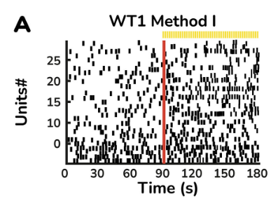
Light-driven spiking activity recorded from presumed ON-Centre retinal ganglion cells (RGCs) and OFF-Centre RGCs. In the raster plot, each small vertical bar indicates the time stamp of a spike, where each row represents a different RGC. The left half illustrates the activity before stimulus onset and separated by the red line, the right half illustrates the activity when exposed to light.
iPSC reprogramming (n=3 vials with 1x10⁶ cells per vial shipped to the customer)
RSDR0000RO
Human
Brightfield imaging. Confirmation of Sendai virus clearance via PCR. Flow cytometry for OCT4 and TRA-1-60. Trilineage differentiation assessment.Karyotyping for genetic stability
iPSCs differentiation to retinal organoids
RSDD0000RO
Human
Brightfield imaging
iPSCs differentiation to retinal organoids
RSDD0000RO
Human
Brightfield imaging. Quantitative IF for VSX2, Recoverin,and SNCG
iPSCs differentiation to retinal organoids
RSDD0000RO
Human
Brightfield imaging. Quantitative IF for Recoverin. RT-PCR for retinal markers
Retinal Toxicity
RST00000RO
Human
Brightfield Imaging. ATP/LDH (basic)
Retinal Toxicity
RST00000RO
Human
Brightfield Imaging, ATP/LDH. Qualitative IF (3 markers). TUNEL (comprehensive)
Retina Disease Modelling
RSD00000RO
Human
Brightfield imaging
Retina Disease Modelling
RSD00000RO
Human
Brightfield imaging. Quantitative IF for VSX2, Recoverin and SNCG
Retina Disease Modelling
RSD00000RO
Human
Brightfield imaging. Quantitative IF for Recoverin. RT-PCR for retinal markers
Retina Gene Therapy Evaluation
RSG00000RO
Human
Brightfield imaging. Fluorescence imaging on live organoids
Retina Gene Therapy Evaluation
RSG00000RO
Human
Brightfield imaging. Fluorescence imaging on live organoids. ATP, LDH and Flow Cytometry for Annexin V for cell viability and apoptosis.Qualitative IF with co-staining for GFP and retinal markers. Flow cytometry for analysing transduction efficiency. RT-PCR for key retina makers
Live Retinal Organoids Product
Human iPSC-derived retinal organoids (n=10) in 5 ml vial filled with organoid culture medium
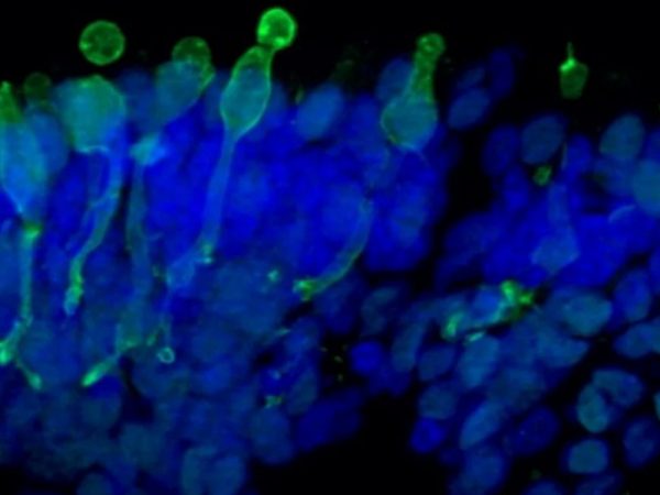
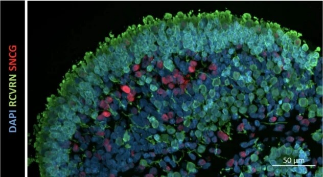
Localization and distribution of photoreceptors (RCVRN, green) and retinal ganglion cells (SNCG, red) in retinal organoids at d150. Nuclear DAPI staining (blue)
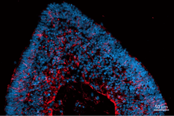
Localization and distribution of Müller glia cells (CRALBP, red) in retinal organoids at d180. Nuclear DAPI staining (blue).
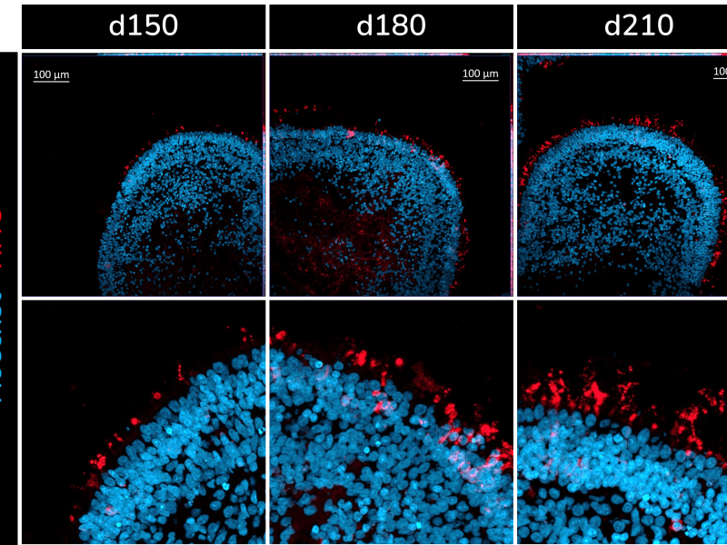
Presence of rod photoreceptors (RHO+) at different stages of development in iPSC-derived retinal organoids. ‘d’ refers to ‘days of differentiation’.
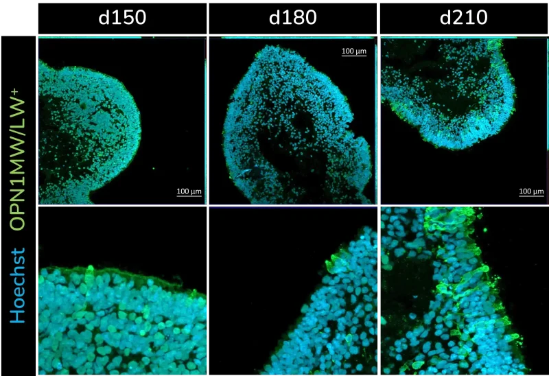
Presence of green and red cone photoreceptors (OPN1MW/LW+) at different stages of development in iPSC-derived retinal organoids. ‘d’ refers to ‘days of differentiation’.