Predictive retinal models for advanced toxicity assays
Accelerate your lead compound selection by understanding their toxicology profile in functional retinal models
1.
Recapitulate the architecture & function of the human retina
2.
Evaluate novel drugs safety profile in vitro
3.
Accelerate drug discovery & replace animal experiments
Predictive in vitro evaluation of retinal toxicity
The retinal drug toxicity in vitro studies are fast, reliable and use the highly reproducible iPSC-derived retinal organoids and/or RPE models. The models recapitulate the native tissues in structure and function and are known to respond to toxins.
The basic retinal toxicity study evaluates how new compounds affect cell morphology and cell viability.
The comprehensive retina toxicity study includes the readouts from the basic study as well as functional assays for RPE and key marker analysis for retinal organoids.
The studies can be carried out at different time points of differentiation and will provide predictive data to allow for rapid decision making to progress your new drug or submit an IND.
Service outputs
Rapid retinal toxicity evaluation of compounds in iPSC-derived retinal organoids and RPE
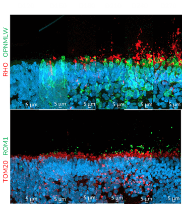
Quantitative and qualitative analysis of key markers expression by gene expression analysis and immunofluorescence on frozen retinal organoid sections
Time point comparisons
Compound effect on PRE barrier, phagocytic integrity, viability and ultrastructure
Newcells provides retinal toxicity studies using our highly reproducible iPSC-derived human models (retinal organoids and RPE) to provide you with predictive data
With a streamlined manufacturing process releasing regular supply of high quality tissue, projects timelines are usually short and a study is usually carried out within two months. The predictive data generated by our scientific experts will therefore allow you to rapidly assess the ocular toxicity profile of your drugs to guide key decision-making steps during drug development or regulatory submissions.
An example of retinal drug toxicity screening includes a set of advanced assays to assess cell viability, photoreceptor functionality and degeneration as well as analysis of key marker expression and localisation to test novel drug compounds and viral vectors such as AAV.
What is retinal toxicity and how can it be evaluated in vitro? Some drugs administered systematically may affect the function of the retina. Similarly, new treatments for eye diseases require careful safety evaluation. Retinal toxicity studies can be performed in animal models, or human ex-vivo models. However, these are limiting due to the number of experimental data points that can be generated and limited predictivity in humans.
Newcells scientists have developed robust advanced retinal toxicity assays run on highly reproducible and readily available iPSC-derived human organoids or RPE models that recapitulate the in vivo environment.
Retinal organoids are obtained through a carefully controlled differentiation process recapitulating the timeline of embryonic retinogenesis. At day 150, the organoids comprise all key cell types and are functional, allowing the detailed evaluation of the cytotoxic effect of new compounds by simply adding them to the plate. The cell structure integrity and the gene expression profiles of key markers for the main cell types, such as photoreceptors, are also assessed following addition of the drug.
Qualitative imaging and microscopy analysis are performed on the models to provide a comprehensive drug toxicity profile.
Uniquely, the RPE cells are derived from healthy donor iPSCs from the same genetic background as retinal organoids, allowing parallel and isogenic assessment of both RPE and neurosensory retina. The plates format of the RPE model allows for flexibility in dosing and analytical readouts in toxicity studies, including morphology assessment of the cells and functional assessment such as RPE-analysis of phagocytosis of photoreceptor outer segments and trans-epithelial resistance (TEER).
As our assays are preformed with models derived from human iPSCs, they are directly relevant to human clinical trials, and provide key toxicology predictive data for translational studies. The two main advantages of outsourcing your in vitro retinal toxicity study are the speed (as most simple studies can be carried out within one to two months) and the data predictivity for human clinical trials.
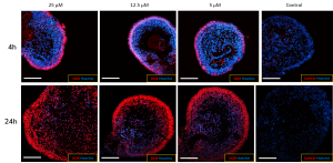
Retinal drug toxicity study in retinal organoids: The response of retinal organoids to exposure of known toxins such as thioridazine and doxorubicin. The intrinsic fluorescence of doxorubicin facilitates visualisation of the drug penetrating the retinal organoid (A). Exposure of the iPSC-derived retinal organoids to doxorubicin reduces cell viability in a dose-dependent manner (B).
(A) Newcells’ human iPSC-derived retinal organoids are permeable to small molecules. The penetration of doxorubicin, a naturally fluorescent small toxic molecule (red), into the retinal organoids increases over time (4h to 24h) demonstrating the permeability of the organoids to drugs.
(B) The retinal organoids were treated with increasing dose of doxorubicin over a period of 24h and cell viability was measured using an ATP assay. A dose-dependent decrease in cell viability was observed following increasing exposure to the drug.
During early drug development, drug screening is an essential step for the identification of lead compounds. This is usually performed as medium to high throughput. Newcells Biotech retinal organoids are suitable for in vitro drug screening since they are generated every 4-6 weeks (on-demand supply) in a 96-well plate format. They have been used to distinguish compounds which are toxic and non-toxic to the retina.
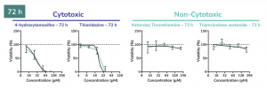
Dose-response plots of known cytotoxic and non-cytotoxic retinal agents. Retinal organoids were exposed to increasing concentrations of either cytotoxic or non-cytotoxic drugs and cell viability was measured over time. As expected, cell viability decreased upon increasing addition of Thioridazine and 4-hydrohytamoxifen, whilst non-cytotoxic drugs had no effect. The dose-response of cytotoxic and non-cytotoxic retinal agents was determined using CellTiter-Glo® 3D ATP assay.
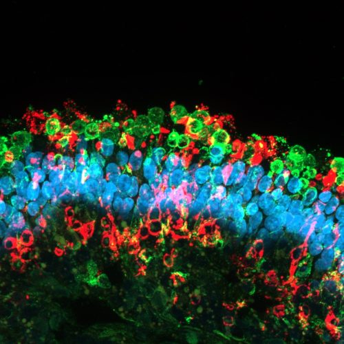
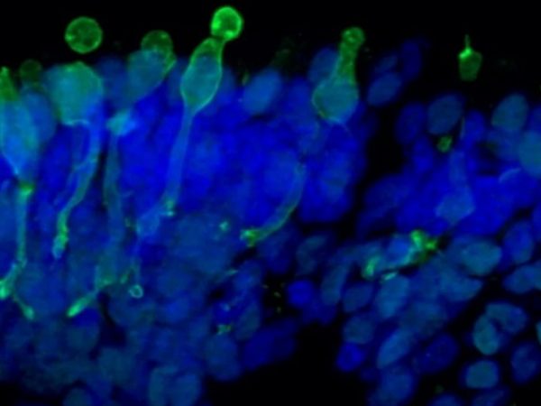
The human retinal organoids are iPSC-derived microtissue models. They recapitulate the complex structure of the human retina with laminar cell organisation mimicking embryonic development. They contain the outer photoreceptor segment of the retina that responds to light.
Cone photoreceptor cells labelled with anti-Opsin (Red/Green) antibody.Find out more
Retinal pigment epithelium
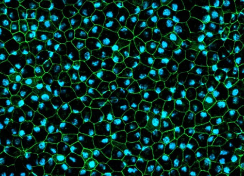
A functional human 2D in vitro model of retinal pigment epithelial cells generated from human iPSCs recapitulating phagocytosis of photoreceptor outer segments. The RPE cells are pigmented and displays typical cobblestone morphology making them a complex human in vitro model able to produce predictive translational data.
RPE cells displaying cobblestone morphology. Cells were immunolabeled with tight-junction marker ZO-1 (shown in green) and co-stained with nuclei marker Hoechst (shown in blue).Read more