Analysis of the effect of a compound on retinal microtissue composition
Tissue
Retina ModelsModels and Services
Retinal Organoids, Retinal Disease Modelling Service, Retina Efficacy Study, Retinal Toxicity ServiceThe Problem of the Client
The client wished to analyse the effect of their compound on the cellular composition of retinal tissue using mature disease model organoids.

Solution
We generated organoids containing the specific disease mutations and quantified cell-type specific biomarkers for horizontal cells, bipolar cells, amacrine cells, rod photoreceptors, Müller glial cells and astrocytes following treatment with the compound.
Step 1:
Understanding the Problem
Through in-depth discussion with the client, we agreed that the study should focus on two specific mutations relevant to the disease of interest and decided to generate retinal organoids from two gene-edited iPSC lines containing these mutations. A set of immunofluorescence-based studies was then needed to quantify each cell population within the organoids.
Step 2:
Developing a Customised Experimental Plan
We designed a study divided into 2 parts and presented the plan in our Statement of Work.
Part 1: Generation of retinal organoids from disease iPSC lines (mutations A and B).
Part 2: Comparison of organoid cell composition in disease organoid models with wild type organoids in the presence and absence of compound X.
The cell types analysed included horizontal cells, bipolar cells, amacrine cells, rod photoreceptors, Müller glial cells and astrocytes. Cell types were identified on frozen sections by a cell specific biomarker expressed at day 180 and day 210 of differentiation. The percentage of cells expressing the biomarker over the total number of cells was determined by quantitative immunofluorescence analysis.
Step 3:
Project Execution
Our scientists carried out the agreed experiments in the proposed timeframe, providing regular updates to the client.
The study included the experimental phase, data processing, data analysis and presentation of a comprehensive data summary.
The experiments were carried out on 10 mm frozen sections of mature day 180 and day 210 retinal organoids and analysed with a Zeiss Axio Apotome fluorescence microscope (20X objective).
Output Data Set:
The complete study delivered a comprehensive dataset comprising of:
- Comparison of Organoid lines: Mutation A, Mutation B, Isogenic WT control, internal WT positive control.
- Comparison of two time points of organoid differentiation: day 180 and day 210 of differentiation
- Number of organoids analysed per experimental condition: 6
- Cells quantified (cell specific marker): horizontal cells (PROX1), bipolar cells (VSX2 and PKCa), amacrine cells (AP2a), rod photoreceptors (RHO), Müller glial cells (CRALBP) and astrocytes (GAFP).
Example data set that was provided to the client:
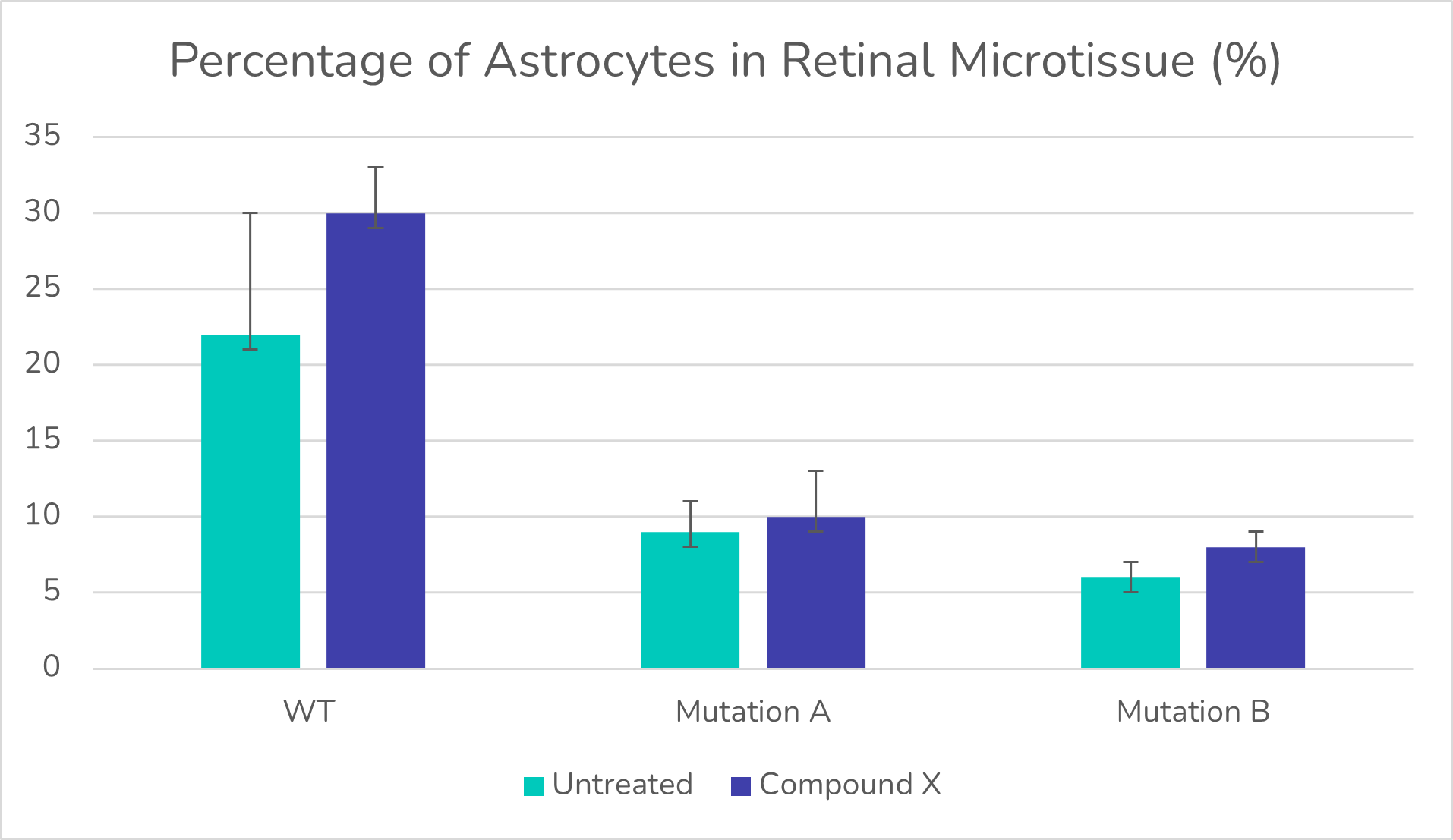
Percentage of Astrocytes in retinal microtissue.
Step 4:
Delivery of Results
We delivered a detailed report of the dataset which was shared digitally and discussed over a call.
The study clearly showed:
- More pronounced differences in retinal tissue cell composition at day 210 compared to day 180 of differentiation.
- Both models of disease retinal organoids displayed a higher proportion of horizontal cells and amacrine cells at day 210 compared to WT.
- Both models of disease retinal organoids displayed a lower percentage of Müller glial cells and astrocytes compared to wild type.
- The percentage of bipolar cells in retinal tissue was not affected by Mutation A or B.
- The addition of compound X increased the percentage of horizontal cells, decreased the fraction of rods photoreceptors and Müller glial cells.
Outcomes for the Client and the Project
The client was able to:
1) Confirm how specific disease mutations affect the composition of retinal tissue in a human in vitro model.
2) Assess the effect of their compound on cellular composition to provide additional safety and efficacy data for their IND submission.
