Why use our renal models?
Express all major renal transporters
- Relevant results
Available to model most pre-clinical species
- Predictive of human outcomes
Extensively validated for DDI and nephrotoxicity
- Reliable data
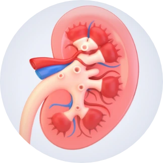
Kidney tissue models
aProximate™
aProximate™ is a validated, near physiological renal transporter model for use in drug transport and renal toxicity studies.
Products
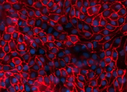
Human Primary Proximal tubule cells
Services
Drug drug interactions and DDI
- Drug transporter assay with primary proximal tubule cells (species : Human, rat, mouse and dog)
- Drug interactions assay with assay with primary proximal tubule cells (species : Human, rat, mouse and dog)
- Flux and net transporter measurements assay with primary proximal tubule cells (species : Human, rat, mouse and dog)
- Measurements of intracellular drug and metabolite concentrations assay with primary proximal tubule cells (species : Human, rat, mouse and dog)
Renal toxicity
Cross-species comparison
Kidney disease modelling
Newcells Offerings
Dr Mike Nicholds
Working with us
Timely service
Our ethical and secure primary tissue supply chain enables us to run studies and produce products to meet customers important and demanding timelines. We process multiple primary tissue every month meaning order-to-data response times can fit with the most demanding of customer deadlines.
Model validation
Our kidney tissue models are well-characterised and rigorously validated to ensure the generation of robust and comparable data predictive of in vivo renal physiology. Our clients also value our scientific expertise supporting the experimental design as well as the analysis and interpretation of data.
How we support our clients
By providing predictive data, we give our clients confidence in their key decision processes using our renal transporter models. Our service enables identification of cross-species differences in renal drug handling, identification of clinically relevant transporter-mediated Drug-Drug interactions and assessment of drug-induced kidney damage.
Our expertise
Our team have an in-depth knowledge of kidney physiology, cell-based models and assay development. They have designed and completed hundreds of studies on novel compounds using the unique renal models and provided interpreted data sets to customers to advance candidates into the clinic. Working under a QMS environment, the data and reports have been used to support regulatory submissions.
