Skip to content
Announcement:

Download our latest eBook on lung airway epithelial cell and fibroblast models

A young man adding liquid to a test tube

Disease Modelling

Renal Transporters and disease

  • Calcium and Phosphate transporters imbalance
  • Amino acid transporter impairment
  • Urate transporters deficiency

Models

  • aProximate™ kidney proximal tubule cells
  • Glomerulus model

Species

  • Human
  • Mouse
  • Rat
  • Dog
  • NHP

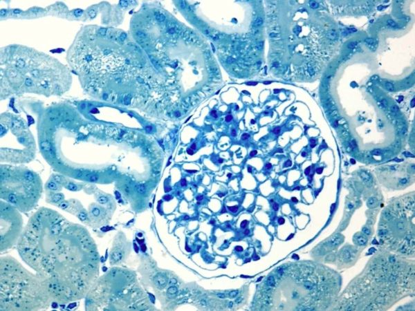
Molecular and cellular mechanisms of kidney disease modelling in vitro

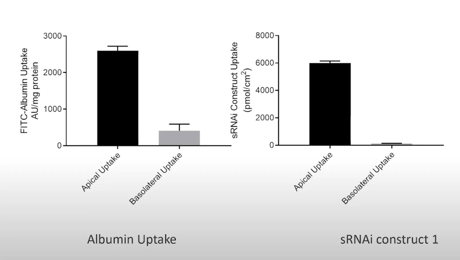
To study renal dysfunction and perform in vitro kidney disease modelling, a robust model, such as aProximate™, expressing all the transporters specialised in handling of drugs, solutes, and proteins is required. The Newcells platform is unique as it is highly predictive of in vivo outcomes. It also allows insights into disease mechanisms and assessment of  therapeutic targets (e.g., renal transporters) of new drugs. aProximate™ has been used to study a range of diseases including chronic kidney disease (CKD), gout, and metabolic disorders.

Service outputs

  • Apical to Basal (Jab) and Basal to Apical (Jba) flux
  • Net transport

Don't miss out on our latest innovations: follow us on Linkedin

In vitro renal tubular drug transport modelling

Chronic kidney disease (CKD)

Metabolic disorders

Gout

Newcells aProximate™ and glomerulus model

Accelerate your lead compound selection by understanding their mode of action in kidney tissue.

1.

Fully differentiated and polarised kidney proximal tubule cells

2.

High expression of all relevant transporters

3.

High predictivity of in vivo and clinical outcomes

Modelling molecular and cellular mechanisms of disease Close Open

The kidneys are essential organs playing a key role in homeostasis and maintaining the right balance between fluids, electrolytes, nutrients, and toxins. Renal dysfunction is either caused by external factors, such as drugs or toxins, or is linked to other underlying conditions affecting the pancreas and the heart, such as diabetes mellitus and cardiovascular disease. Rare inherited metabolic disorders also affect vital organs including the kidney. The kidneys ensure waste is excreted from the blood and nutrients are reabsorbed from the urine; a function performed by the kidney proximal tubule cells through a set of sophisticated transporters expressed both at the apical and basolateral cell membranes. When this equilibrium is perturbed, kidney function is affected.

Example 1: Chronic Kidney Disease (CKD) – calcium and phosphate transport balance Close Open

CKD is defined as a change in kidney function or structure affecting the health of an individual for longer than 3 months irrespective of the cause. The early stages of CKD are asymptomatic, whilst the end-stages are treated by dialysis and kidney transplant. The key cellular and molecular events causing CKD are an imbalance in the renin-angiotensin-aldosterone system, chronic inflammation, fibrosis, the presence of uremic toxins, and vascular calcification. Vascular calcification is not unique to CKD, but is also seen in diabetes mellitus and cardiovascular disease, other factors associated with CKD. Vascular calcification occurs due to an imbalance in calcium and phosphate homeostasis, often as a result of a decrease in renal phosphate clearance. Renal sodium and phosphate co-transporters such as sodium-phosphate transporters type I and II (NaPi-I and NaPi-II) are therapeutic targets for CKD. These and other transporters are expressed in aProximate™, making it an ideal tool to model CKD in vitro by measuring the rate of intracellular accumulation of phosphate.

Example 2: Metabolic disorders – impairment of renal tubular transport of amino acids Close Open

There is a group of inherited diseases with inborn errors of metabolism where, for example, the transport of solutes such as salts and amino acids across the apical membrane of proximal tubule cells is affected. As a result, the reabsorption of essential amino acids is impaired, leading to an excess of urinary excretion and amino acid loss from the body. The most frequent disorders are caused by mutations in solute carrier membrane transport proteins (e.g., SLC3, SLC6, SLC7, SLC15). These mutations are present in metabolic disorders such as Iminoglycinuria, Cystinuria, Fanconi Syndrome, Nephrolithiasis, Hartnup disorder and Gitelman Syndrome.

aProximate™ proximal tubule cells express transporters of the SLC family (SLC3, SLC6, SLC7, SLC15) allowing the uptake of essential amino acids by PTCs to be measured and modelled with this unique in vitro model. High throughput screening of drug modulators such as amino acid transporters is also possible with our aProximate™ PTC models.

Example 3: Gout – uric acid transport Close Open

Uric acid is a waste product transported and eliminated by the kidney. When produced in excess, the kidneys are unable to excrete it fast enough and it accumulates in the blood (hyperuricaemia). This condition is known as gout and there is currently no effective clinical treatment for it. High levels of uric acid in the blood are the hallmark of gout, and sometimes also a sign of CKD. Urate transporters, such as NPT1, NPT4, OAT1, OAT2, OAT3, OAT4, URAT1, GLUT9, ABCG2 and PDZK1 have been shown to play a role in the upregulation of serum uric acid levels, making them potential therapeutic targets for new treatments of gout. Targeting urate transporters and investigating urate-lowering drugs in vitro can provide insights into hyperuricaemia and related disorders.

With urate transporters expressed in aProximate™ PTCs, in vitro high throughput analysis of inhibitors is straightforward. Given that aProximate™ is also highly predictive of in vivo outcomes, it is an ideal in vitro tool to understand uric acid transport and evaluate new treatments for gout.

Example 4: Therapeutic Oligonucleotide and siRNA delivery to the proximal tubule Close Open

Megalin and Cubilin are key uptake transporters of siRNA and oligonucleotides in the proximal tubule. They also transport large molecules such as albumin, insulin and aminoglycosides. aProximate™ proximal tubule cells express functional levels of Megalin and Cubilin, allowing the testing of siRNA uptake by PTCs with this unique in vitro model.


siRNA and albumin uptake experiments in freshly isolated primary proximal tubule cells

Table
Models

aProximate™ primary isolated kidney proximal tubule cells

Species
  • Human
  • Rat
  • Dog
  • Non-human primate

Model available for this service

aProximate™ proximal tubule cells

aProximate™ is one of the most advanced, near physiological, in vitro, kidney proximal tubule cell (PTCs) models. aProximate™ PTCs are derived from fresh human, rat, mouse, dog and NHP kidney tissue and grown on Transwells® where they remain well differentiated as a polarised cell layer that forms tight junctions.

More information
A microscope image of a nephron model

Glomerulus model

The first fully differentiated, primary podocyte model derived from human kidney tissue. The effect of a drug on the glomerular filtration barrier and the podocyte glomerular permeability can now be easily evaluated in vitro. Podocyte injury and proteinuria can be modelled in vitro at high throughput and assessed by measuring podocyte damage biomarkers, TEER and podocyte permeability.

More Information
A microscope image of a convoluted tubules

Contact us

Is this the service for you? Speak to one of our experts about your requirements.

Make an enquiry

Get our latest updates straight to your inbox