
Resources: Posters
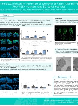
Generation of a physiologically relevant in vitro model of autosomal dominant Retinitis Pigmentosa caused by RHO-P23H mutation using 3D retinal organoids
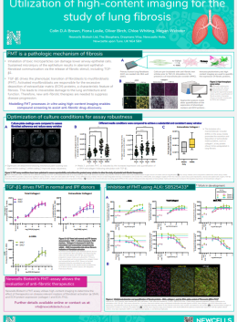
Utilization of high – content imaging for the study of lung fibrosis
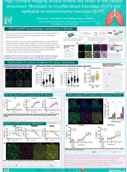
High content imaging (HCI) assays enable the study of the fibrotic processes: fibroblast – to – myofibroblast transition (FMT) and epithelial – to – mesenchymal transition (EMT)
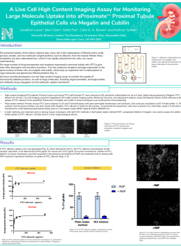
The aProximateTM model for renal drug safety/efficacy and transporter studies
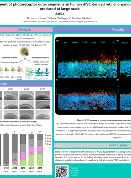
Development of photoreceptor outer segments in human iPSC derived retinal organoids produced at large scale
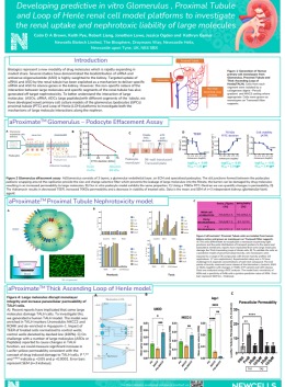
Developing predictive in vitro Glomerulus , Proximal Tubule and Loop of Henle renal cell model platforms to investigate the renal uptake and nephrotoxic liability of large molecules
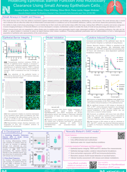
Modelling Epithelial Barrier Function and Mucociliary Clearance Using Small Airway Epithelium Cells
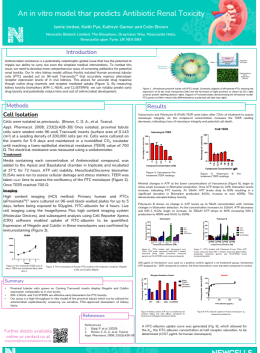
An in vitro model that predicts Antibiotic Renal Toxicity
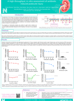
A high-throughput, in vitro assessment of antibiotic induced podocyte injury


