Functional, validated commercially-licensed human cell-based models of the retina to study efficacy and safety of innovative ophthalmology therapies
Models available
- Neural retina (fully stratified including photoreceptors) at Day 150
- Neural retina (partially differentiated) at Day 60
- Day 150+ available on request
- Retinal pigment epithelium (RPE)
- Healthy donor
- Disease models
- Neural retina (fully stratified including photoreceptors) at Day 150
- Neural retina (partially differentiated) at Day 60
- Day 150+ available on request
- Retinal pigment epithelium (RPE)
- Healthy donor
- Disease models
Assays Available
- Drug permeation
- Dose response to drugs
- Organoid viability and cytotoxicity assays
- Gene expression & transcriptomics
- Flow cytometry & Immunofluorescence
- Electron-microscopy
- Drug permeation
- Dose response to drugs
- Organoid viability and cytotoxicity assays
- Gene expression & transcriptomics
- Flow cytometry & Immunofluorescence
- Electron-microscopy
Tissue types
- Ethically sourced healthy human
- Diseased human (retinopathies)
- Ethically sourced healthy human
- Diseased human (retinopathies)
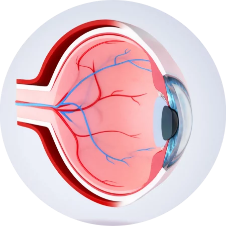
Retina tissue models
Retinal Organoids
Functional iPSC-derived retinal organoids with all the cell types and laminar cell organisation that recapitulate the complex structure of the human retina. They contain the outer photoreceptor segment of the retina that responds to light.
More information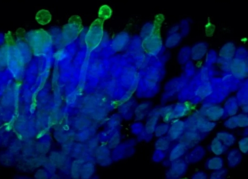
Retinal Pigment Epithelium (RPE)
A functional 2D model of retinal pigment epithelial cells generated from human iPSCs recapitulating phagocytosis of photoreceptor outer segments. The RPE cells are pigmented and displays typical cobblestone morphology.
More information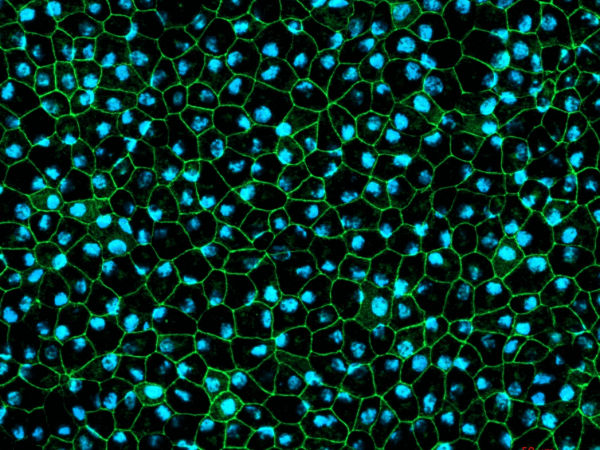
Using validated retina tissue models
Accurately mimics physiology of the human retina
No donor variation
Short timelines to results
Services
Disease Modelling
We ethically source patient lines or use gene editing to produce models of retinopathies.
Retinal toxicity evaluation
High throughput in vitro drug screening for retinal safety testing in a human relevant model.
Gene therapy
Rapid in vitro preclinical evaluation of viral vectors for retinal therapy development.
Working with us
Timely service
With regular manufacturing of retinal organoid batches, our services and product supply timelines are short. The retinal organoid can be harvested at various times points during differentiation according to the maturation stage of the main cell type of interest. Typically, we offer organoids at day 150, but we offer full customisation of the model to suit your requirements.
Model validation
Our retinal organoids and RPE models are well-characterised and rigorously validated to ensure the generation of robust and comparative data predictive of the human retina. Assays have been optimised to work with the unique nature of 3D retinal organoids.
How we support our clients
By providing predictive data, we give our clients confidence in their key decision processes, such as the selection of the optimal viral vector for gene therapy or selection of a lead compound in vitro for retinopathies.
Our expertise
Investing over 3 years in development and completing numerous studies for customers our team has unrivalled expertise in application of human iPSC models of the retina. We can work with you to design the right study to provide data on large and small molecule effects on the retina to assist in candidate progression.
