Skip to content
Announcement:

Our latest paper in collaboration with Pfizer on the role of OCT2/MATE in creatinine clearance is accepted in JPET.

Liquid being dropped into a test tube

Retinal Organoids

Model formats

  • Fresh retinal organoids
  • Retinal organoid cell pellets
  • Total RNA preparation of retinal organoids
  • Frozen sections of retinal organoids
  • Available at various customisable time points e.g. D60, D150, D180, D210

Cell Types

  • Rod and cone photoreceptors
  • Retinal ganglion cells
  • Bipolar cells
  • Horizontal cells
  • Amacrine cells
  • Müller glial cells

Species

  • Human (Healthy donor)
  • Human (Gene-edited)

Light responsive retinal organoids for accurate prediction of clinical outcomes

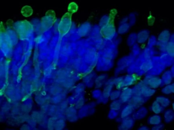
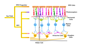
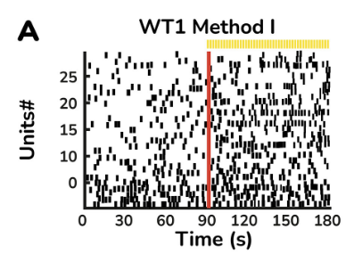
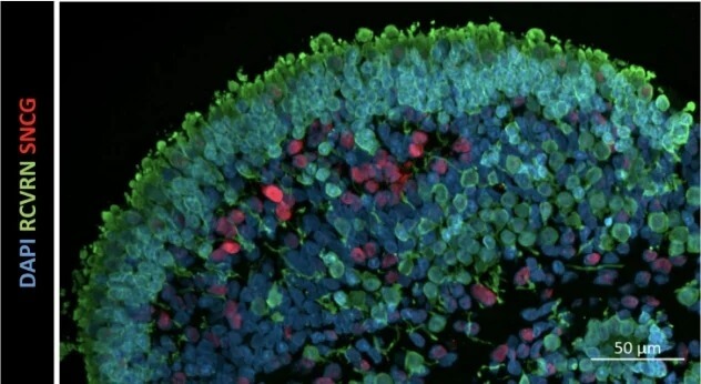
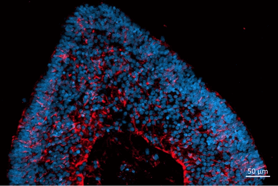
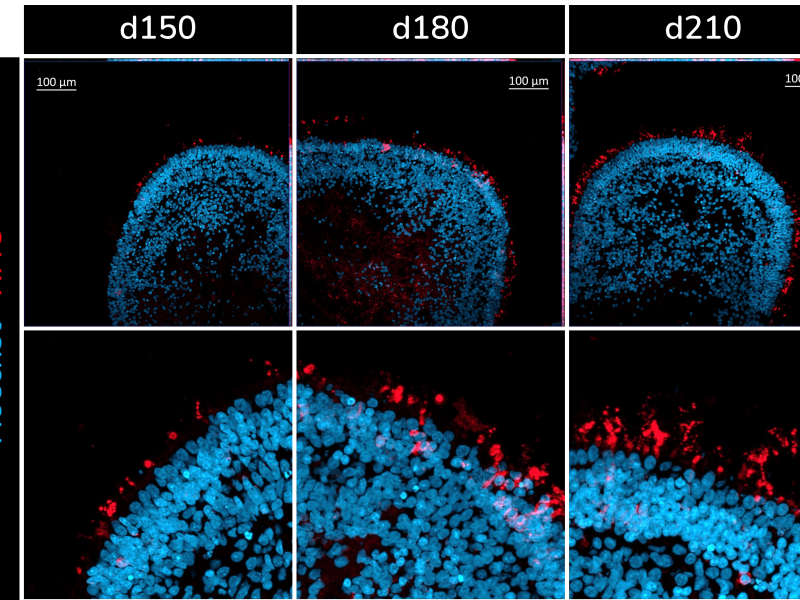
The retinal organoids recapitulate the complex structure of the human retina with laminar cell organisation mimicking embryonic development. They contain the outer photoreceptor segment of the retina that responds to light.

Available analytical readouts

  • Immunofluorescence analyses
  • mRNA quantification by RT-qPCR
  • Transcriptomic analysis by single-cell RNA sequencing
  • Cytotoxicity assays
  • Cytokine release
  • Flow cytometry
  • Electron microscopy
Make an enquiry

Don't miss out on our latest innovations: follow us on Linkedin

A microscope image of retinal organoids
Cone photoreceptor cells labelled with anti-Opsin (Red/Green) antibody.

Origin

Healthy donor or gene edited

Multiple cell types

3D structure

Lead Time

4-6 Weeks

Retinal Organoid Model Highlights

Accelerate your lead compound selection by understanding their mode of action in functional retinal tissue

1.

Complex functional human retina model

2.

Well-characterised and reproducible

3.

Ready-to-use

Newcells Retinal Organoids

Hannah Steward, Associate Scientist

Applications for retinal organoids Close Open
  1. Gene therapy, including in vitro viral vector evaluation
  2. Disease modelling
  3. Investigational drug safety and efficacy
Retinal Organoid Characterisation Close Open

In vitro retinal organoid differentiation follows the embryonic development timeline, spanning 150 to 210 days. The temporal order of retinogenesis is comparable to in vivo, recapitulating critical features of foetal retinal tissue including the laminar organisation of cell types.

The organoids include retinal ganglion cells, horizontal cells, amacrine cells and photoreceptors (including cone and rod photoreceptors). Depending on the cell type of interest, the retinal organoids can be used at different stages of development (usually between D60 and D210). For example, retinal ganglion cells are more prevalent at D60 whereas the photoreceptors have a peak expression at later time points.

Retina illustration
Schematic representation of the retina

The differentiation process is fully characterised and monitored through the analysis of biomarkers specific for each cell type. For example, for cone photoreceptors, we monitor the expression of OPNSW, OPNMW, OPNLW, ARR3, RXRG; for rod photoreceptors we use RHO and NRL and for retinal ganglion cells we follow MATH5 (ATOH7) and BRN3 (POU4F2). Our organoids are validated for many applications.

Cell type Gene Timepoint of appearance Timepoint of peak expression
Retinal ganglion cells MATH5 (ATOH7) d30 - d180 d60
BRN3 (POU4F2) d30 - d210 d60
Horizontal and amacrine cells TFAP2A d30 - d210 d150
PROX1 d30 - d210 d150
Bipolar cells GRIK1 d30 - d210 d150
CADPS d30 - d210 d150
Photoreceptors RCVRN d60 - d210 d210
RBP3 d60 - d210 d210
IMPG1 d120 - d210 d210
CRX d60 - d210 d210
Cone photoreceptors OPN1SW d120 - d210 d210
OPN1MW d150 - d210 d210
OPN1LW d120 - d210 d210
ARR3 d60 - d210 d180
RXRG d60 - d210 d150
Rod photoreceptors RHO d120 - d210 d210
NRL d90 - d210 d180
RPE RPE65 d60 - d210 d210
Muller glia RLBP1 d90 - d210 d210
CRYM d30 - d210 d210
Cell type Cell marker Protein expression at d150 Protein expression at d180 Protein localisation at d150-d180
Photoreceptors RCVRN ONL
Retinal ganglion cells SNCG and Huc/D INL/GCL
Cone photoreceptors OPN1MW/LW ✓ * ONL
Rod photoreceptors RHO ✓ * ONL
Bipolar cells PKC-α ** INL
Amacrine cells AP-2α INL
Horizontal cells PROX1 INL
Muller glia CRALBP All layers

*Small number of developing rods and cones

**Expressed at transcriptional level

Retinal organoids description Close Open

Characteristics of iPSC-derived retinal organoids:

  1. Size: ~1.3 mm in diameter
  2. Number of cells: ~ 40,000
  3. Cell types: retinal ganglion cells, horizontal cells, amacrine cell and photoreceptors (including cone and rod photoreceptors)
  4. Structure: fully-stratified, similar to the human retina
  5. Main characteristics: formation of primitive photoreceptor outer segments, recapitulate retinogenesis in vitro
  6. Other characteristics: responsive to known toxins, functional and responsive to light, all cell layers allow drug permeation
Light response of the retinal organoids
Light-driven spiking activity recorded from presumed ON-Centre retinal ganglion cells (RGCs) and OFF-Centre RGCs. In the raster plot, each small vertical bar indicates the time stamp of a spike, where each row represents a different RGC. The left half illustrates the activity before stimulus onset and separated by the red line, the right half illustrates the activity when exposed to light.
Retinal organoids kit content Close Open
  • 10 fresh organoids per vial
  • Optimized cell culture medium
  • 96 well plate
  • Pasteur pipette

Other formats available

  • Fresh retinal organoids
  • Retinal organoid cell pellets
  • Total RNA preparation of retinal organoids
  • Frozen sections of retinal organoids (10μm) for immunofluorescence staining

All retinal organoids are available at various customisable time points: D60, D150, D180, D210

Images

DAPI RCVRN SNCG
Localization and distribution of photoreceptors (RCVRN, green) and retinal ganglion cells (SNCG, red) in retinal organoids at d150. Nuclear DAPI staining (blue)
Localization and distribution of Müller glia cells (CRALBP, red) in retinal organoids. Nuclear DAPI staining (blue).
Localization and distribution of Müller glia cells (CRALBP, red) in retinal organoids at d180. Nuclear DAPI staining (blue).
Rod photoreceptors
Presence of rod photoreceptors (RHO+) at different stages of development in iPSC-derived retinal organoids. ‘d’ refers to ‘days of differentiation’.
Cone photoreceptors
Presence of green and red cone photoreceptors (OPN1MW/LW+) at different stages of development in iPSC-derived retinal organoids. ‘d’ refers to ‘days of differentiation’.

Contact us

Interested in our retinal organoids? Speak to one of our experts about your requirements.

Make an enquiry

Get our latest updates straight to your inbox